Benign Excisional Surgery
Excisional Surgery of Benign Lesions
Excision of skin lesions usually involves the use of relatively superficial structures. It is performed under local anesthesia and seldom entails significant blood loss.
The technique is commonly used by physicians to remove skin and subcutaneous lesions. A properly designed elliptical excision has a length-to-width ratio of 3 to 1 and produces a 30-degree angulation at both corners of the wound. After administration of local anesthetic, the lesion is excised and sutures are used for the closure. The skin edges are brought together during the closure in a special way to improve the final scar appearance. Dr. Shvartzman has performed over 90,000 surgical cases and has significant experience producing good cosmetic outcomes.
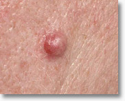
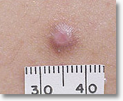
Cysts are extremely common, forming when dead skin plus the oily secretions of the dead skin cells, which usually shed monthly, get trapped under a clogged pore.
Surgical excision of a sebaceous cyst is a simple procedure to completely remove the sac and its contents. With surgery, a cyst can usually be excised in its entirety. A completely removed cyst will not recur. Although, if the patient has a predisposition to cyst formation, they may develop more cysts in the future.
The most commonly excised nevi (moles) include:
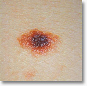
Dysplastic Nevus
The term dysplastic nevus is often used interchangeably with “atypical” mole, although many consider the latter to be a clinical term, with “dysplastic nevus” used to describe certain histologic (microscopic) features of the mole. Pathologically diagnosed dysplastic nevi are often treated by complete removal with a shave or surgical excision.
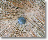
Blue Nevus
Blue nevi are characterized by a collection of pigment-producing melanocytes in the dermis. These lesions are usually bluish or bluish-black colored papules. They are usually benign and stable over time. However, malignant melanomas developing in or associated with a blue nevus have been rarely reported. Due to this, blue nevi are often surgically removed.
Congenital and Interadermal Nevus
Flesh-colored or “brownish” moles. These moles are benign, and are removed for aesthetic purposes or due to irritation. They are usually surgially excised or shaved off to prevent recurring pigmentation.
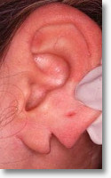
Excessive weight or trauma (especially when an ear is pierced in the wrong location) can overcome the strength of the earlobes, causing a tear in the tissue. This split may be unattractive and renders most jewelry unusable.
Most torn earlobes can be effectively repaired surgically. The procedure is performed with local anesthetic. The tear in the earlobe is excised and the earlobe is cosmetically repaired resulting in a straight line of suturess on the front and the back of the earlobe.
The ear can be repierced after the scar heals. Dr. Shvartzman will let you know when you can safely re-pierce your ears, and advise you on the correct location for re-piercing.
This procedure can be modified to correct large defects in the ear from gauges.
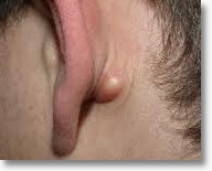
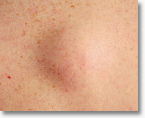
A lipoma is a fatty tumor composed of adipose (fat) tissue, and is usually benign. Typically, these tumors take the form of a lump that forms just under the surface of the skin. Lipomas are usually removed when the tumor becomes painful, tender, or enlarges.
Lipomas are best removed through a small incision made in the skin overlying the lipoma, removing the fatty tumor, and closing the wound.
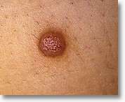
Dermatofibromas are harmless, very common firm bumps that occur in the skin. They range from flesh-colored to dark-brown, with a slightly lighter center than the outside edge. The cause of dermatofibromas is unknown, but some have suggested they may be a scar-like skin reaction to a bug bite or minor injury.If a dermatofibroma occurs in a cosmetically sensitive area or is tender, irritated, or symptomatic, it can be surgically removed.
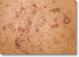
Neurofibromas are common benign lesions derived from cells that surround nerves. They can occur anywhere on the body and may be single or multiple. Multiple neurofibromas are often a sign of neurofibromatosis, an inherited disorder which affects the skin, bone and nervous system.
The lesions are single or multiple soft, flesh colored, tan or pink bumps which may be on a stalk. The lesions can be pushed down through the skin with pressure. This phenomenon is known as “button-holing”.
Most large neurofibromas require surgical removal. Surgical removal allows the patient to have minimal wound care and a better cosmetic outcome.